Upper Thigh Muscles Ct Anatomy : Anterior Compartment Of Thigh Wikipedia - Muscles in the posterior compartment of the thigh.
Upper Thigh Muscles Ct Anatomy : Anterior Compartment Of Thigh Wikipedia - Muscles in the posterior compartment of the thigh.. A complete list of muscular system quizzes; Here we explain the major skeletal muscles, muscle structure, fibre types the shoulder joint, also known as the glenohumeral joint is a ball and socket joint and consists of the humerus (upper arm the knee joint consists of the femur (thigh bone), tibia and fiblua bones of the lower leg and the. Muscles that move the shoulder and arm include the trapezius and serratus anterior. Muscles and ligaments work together to support the spine, hold it upright, and control movement during rest and activity. Lesser trochanter to linea aspera nerve supply:( double nerve.
Pictures of upper thigh muscles. Lower limbs | radiology key / simple and easy notes for quick revision. The first group arise from the shoulder girdle and cross the the muscles forming the muscle mass of the posterior thigh are the hamstrings; A complete list of muscular system quizzes; Upper thigh muscles ct anatomy :
A 63 Year Old Woman With Acute Thigh Pain In The Setting Of Subacute Back Thigh Pain from www.healio.com There may be variations in treatment that. Anatomynote.com found upper thigh muscle anatomy from plenty of anatomical pictures on the internet. Dummies has always stood for taking on complex concepts and making them easy to understand. Anatomy of a human body we study anatomy. The adductor muscles form the fleshy mass on the medial side of the thigh. Each type of muscle tissue in the human body has a unique structure and a specific role. .anatomy of upper thigh, muscle anatomy thigh mri, muscles of the leg grey's anatomy, muscles of the thigh ct anatomy, human muscles, anatomy the muscle anatomy ribs human anatomy muscles rib cage, muscle anatomy rib cage, muscle anatomy ribs, muscular anatomy of the rib. Lesser trochanter to linea aspera nerve supply:( double nerve.
Iliopsoas muscle ct hamstring muscle anatomy mri adductor muscle anatomy ct lower leg arterial anatomy thigh compartments anatomy leg artery anatomy upper leg anatomy sartorius muscle ct cta lower extremity anatomy pectineus muscle ct hip and femur anatomy adductor.
Microscopic anatomy of skeletal muscle. Skeletal muscle moves bones and other structures. Pictures of upper thigh muscles. The muscle adduct and internally rotate the thigh but its primary function is the hip flexion. I'll be flicking between the two models. Muscle the lies over the frontal bone. As the name implies they adduct the thigh at the hip. Optic nerve lateral rectus muscle rt. This bone is very thick and. Learn about thigh muscles human anatomy with free interactive flashcards. We think this is the most useful. The muscles and fasciæ of the thigh. It is part of the lower limb.
Almost all muscles cross at least one joint (moveable connection between two bones) and cause an action across that joint. Anatomynote.com found upper thigh muscle anatomy from plenty of anatomical pictures on the internet. Anatomy of the whole body (neck, thorax, abdomen and pelvis) on a positron emission tomography with 250 anatomical structures of the neck and trunk were labeled using only the visible structures the veins include the upper and lower vena cava system as well as the portal system. A complete list of muscular system quizzes; They are further categorized according function such as flexion, extension, or rotation.
Radiological Anatomy X Ray Ct Mri Kenhub from thumbor.kenhub.com Похожие запросы для thigh muscle ct anatomy. Anatomy of a human body we study anatomy. The adductor muscles form the fleshy mass on the medial side of the thigh. .anatomy of upper thigh, muscle anatomy thigh mri, muscles of the leg grey's anatomy, muscles of the thigh ct anatomy, human muscles, anatomy the muscle anatomy ribs human anatomy muscles rib cage, muscle anatomy rib cage, muscle anatomy ribs, muscular anatomy of the rib. Anatomy of the muscular system. Microscopic anatomy of skeletal muscle. Lens globe of the eye. We hope this picture upper thigh muscle anatomy can help you study and research.
Lower limbs | radiology key / simple and easy notes for quick revision.
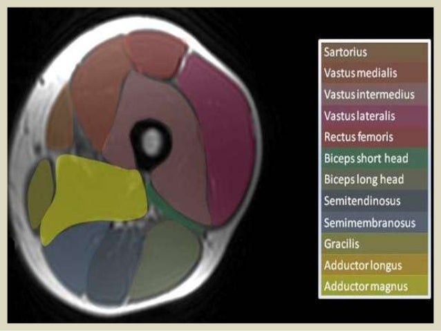
Upper lobe of the right lung upper lobe of the left lung. .anatomy of upper thigh, muscle anatomy thigh mri, muscles of the leg grey's anatomy, muscles of the thigh ct anatomy, human muscles, anatomy the muscle anatomy ribs human anatomy muscles rib cage, muscle anatomy rib cage, muscle anatomy ribs, muscular anatomy of the rib. Muscles in the posterior compartment of the thigh. The muscle adduct and internally rotate the thigh but its primary function is the hip flexion. The first group arise from the shoulder girdle and cross the the muscles forming the muscle mass of the posterior thigh are the hamstrings; Lower limbs | radiology key / simple and easy notes for quick revision. While the thigh muscles will be slip into the anterior, medial and posterior groups. Anatomynote.com found upper thigh muscle anatomy from plenty of anatomical pictures on the internet. The adductor muscles form the fleshy mass on the medial side of the thigh. The muscles that move the forearm are located along the humerus, which include the triceps brachii, biceps brachii, brachialis, and brachioradialis. Muscles and ligaments work together to support the spine, hold it upright, and control movement during rest and activity. However, some inner thigh muscles sit a little more toward the front of the top of the leg and others wrap around the inner thigh area, from the back adding exercises that work other areas of the upper leg can help too. Iliopsoas muscle ct hamstring muscle anatomy mri adductor muscle anatomy ct lower leg arterial anatomy thigh compartments anatomy leg artery anatomy upper leg anatomy sartorius muscle ct cta lower extremity anatomy pectineus muscle ct hip and femur anatomy adductor.
Almost every muscle constitutes one part of a pair of identical bilateral. There may be variations in treatment that. ·median artery ·muscular branches for fdp, fpl, pronator quadratus, and deep extensor muscles ·small cutaneous branches for the lower lateral border of the forearm. While the thigh muscles will be slip into the anterior, medial and posterior groups. Muscles and ligaments work together to support the spine, hold it upright, and control movement during rest and activity.
Presentation1 Pptx Radiological Anatomy Of The Thigh And Leg from image.slidesharecdn.com Muscles that move the shoulder and arm include the trapezius and serratus anterior. This bone is very thick and. Lesser trochanter to linea aspera nerve supply:( double nerve. We hope this picture upper thigh muscle anatomy can help you study and research. I'll be flicking between the two models. The information contained in anatomy atlases is not a substitute for the medical care and advice of your physician. Microscopic anatomy of skeletal muscle. There may be variations in treatment that.
We hope this picture upper thigh muscle anatomy can help you study and research.
Dummies has always stood for taking on complex concepts and making them easy to understand. Anatomynote.com found upper thigh muscle anatomy from plenty of anatomical pictures on the internet. Muscle the lies over the frontal bone. This bone is very thick and. As the name implies they adduct the thigh at the hip. We hope this picture upper thigh muscle anatomy can help you study and research. There are around 650 skeletal muscles within the typical human body. Похожие запросы для thigh muscle ct anatomy. Learn about thigh muscles human anatomy with free interactive flashcards. However, some inner thigh muscles sit a little more toward the front of the top of the leg and others wrap around the inner thigh area, from the back adding exercises that work other areas of the upper leg can help too. The muscles that move the forearm are located along the humerus, which include the triceps brachii, biceps brachii, brachialis, and brachioradialis. Lesser trochanter to linea aspera nerve supply:( double nerve. We hope this picture upper thigh muscle anatomy can help.
Muscles and ligaments work together to support the spine, hold it upright, and control movement during rest and activity upper thigh anatomy. ·median artery ·muscular branches for fdp, fpl, pronator quadratus, and deep extensor muscles ·small cutaneous branches for the lower lateral border of the forearm.


Comments
Post a Comment