Tendon Diagram Under Microscope - Pathological Features Of The Tendon Affected By Tendinopathy Under Download Scientific Diagram : The human thyroid gland functions explained, including cellular level images captured under the microscope with diagrams explaining the different cells.
Tendon Diagram Under Microscope - Pathological Features Of The Tendon Affected By Tendinopathy Under Download Scientific Diagram : The human thyroid gland functions explained, including cellular level images captured under the microscope with diagrams explaining the different cells.. Tenocytes constantly repair small amounts of damage to the matrix under normal circumstances; However, tendon cell activity during growth and homeostatic maintenance is less well defined. Mnemonics that can be used to remember the anatomy of the ankle tendons from anterior to posterior as they pass posteriorly to the medial malleolus of the tibia under the flexor retinaculum in the tarsal tunnel include: They appear as rod shaped dark stained bodies during the metaphase stage of mitosis when cells are stained with a suitable basic dye and viewed under a light microscope. They are actually heavily composed of connective.
They appear as rod shaped dark stained bodies during the metaphase stage of mitosis when cells are stained with a suitable basic dye and viewed under a light microscope. Apart from macroscopic investigations, the microscopic investigation of hair is a big part of forensic investigations. Select the lowest power objective lens. A cell is a very tiny structure which exists in living bodies. The primary function of areolar connective tissue is to give nourishment and cushion to the epithelia.
Definition Of Microscopic Structure Of Muscle Chegg Com from media.cheggcdn.com How to use a microscope. Select the lowest power objective lens. This study explores the interface between dynamic loading and tendon healing across multiple length scales using living tendon explants. Be careful pushing it under the clips that the cover slide doesn't move or crack. Tendons and muscles work together to move bones. Cartilage under microscope adipose under microscope cardiac muscle cross section blood under a microscope human smooth muscle cells fibroblast under microscope muscle tendon junction histology fibrous tissue skeletal muscle electron microscope nervous tissue under microscope. It projects an enlarged and illuminated image o the object to. The primary function of areolar connective tissue is to give nourishment and cushion to the epithelia.
Microscope, instrument that produces enlarged images of small objects, allowing the observer an exceedingly close view of minute structures at a scale microscope slides are small rectangles of transparent glass or plastic, on which a specimen can rest so it can be examined under a microscope.
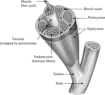
Which is the outer covering of the blood vessel including the esophagus, fascia between muscles, pericardial sacs, and other organs of the body. Materials to be viewed under an electron microscope may require processing to produce a suitable sample. Figure 1.2 light microscope and its parts. Diagram of a transmission electron microscope. They appear as rod shaped dark stained bodies during the metaphase stage of mitosis when cells are stained with a suitable basic dye and viewed under a light microscope. But at the same time it is interpretive. Move the stage (the flat ledge the slide sits on) down to its lowest position. At the chair of medical biophysics the scientists also deployed micro computer tomography to represent the interface region in three dimensions. Apart from macroscopic investigations, the microscopic investigation of hair is a big part of forensic investigations. Start studying anatomy tissues under microscope. Be careful pushing it under the clips that the cover slide doesn't move or crack. Chromosomes were first described by strasburger (1815), and the term 'chromosome' was first used by waldeyer in 1888. Parts of light microscope (fig.
Light microscope t e light microscope is so called because it employs visible light to detect small objects. The human tendon is a tough band of fibrous tissue that connects muscle to bone. Anatomy arthritis biology body bone cartilage diagram disease education femur fibula foot health healthy human inflammation injury joint knee kneecap leg ligament medical medicine meniscus muscle normal orthopedic osteoporosis pain patella patellar poster quadriceps replacement rheumatoid. Related online courses on physioplus. Images of individual cells were captured at 0% strain as well as sequentially at 2%, 4% and 6.
100 Histology Muscle Ideas Muscle Skeletal Muscle Histology Slides from i.pinimg.com Images of individual cells were captured at 0% strain as well as sequentially at 2%, 4% and 6. Move the stage (the flat ledge the slide sits on) down to its lowest position. This video takes you through microscope images of cells going through mitosis and identifies the different phases under the microscope and on a micrograph. The human thyroid gland functions explained, including cellular level images captured under the microscope with diagrams explaining the different cells. We cannot see the structure of a virus under a light microscope because it's size is below the resolution capacity of a classical light. Otherwise, all tendons would weaken and rupture (ker, 2002). The primary function of areolar connective tissue is to give nourishment and cushion to the epithelia. In their relaxed state, the collagen fibers of both tendons and ligaments form a typical wavy pattern, also referred to as a 'crimp,' when viewed under a polarized light microscope.
Similar to our results, chen et.
Which is the outer covering of the blood vessel including the esophagus, fascia between muscles, pericardial sacs, and other organs of the body. Figure 1.2 light microscope and its parts. Start studying anatomy tissues under microscope. The human thyroid gland functions explained, including cellular level images captured under the microscope with diagrams explaining the different cells. The primary function of areolar connective tissue is to give nourishment and cushion to the epithelia. A cell is a very tiny structure which exists in living bodies. The human thyroid gland functions explained, including cellular level images captured under the microscope with diagrams explaining the different cells. They are actually heavily composed of connective. Parts of light microscope (fig. Similar to our results, chen et. Materials to be viewed under an electron microscope may require processing to produce a suitable sample. Select the lowest power objective lens. In their relaxed state, the collagen fibers of both tendons and ligaments form a typical wavy pattern, also referred to as a 'crimp,' when viewed under a polarized light microscope.
Diagram of a transmission electron microscope. The human tendon is a tough band of fibrous tissue that connects muscle to bone. Mnemonics that can be used to remember the anatomy of the ankle tendons from anterior to posterior as they pass posteriorly to the medial malleolus of the tibia under the flexor retinaculum in the tarsal tunnel include: Materials to be viewed under an electron microscope may require processing to produce a suitable sample. Appendix images under the microscope captured with an hd microscopy camera and info on the appendix.
Tendon Slide Wsu 1 041 from undergraduate.vetmed.wsu.edu Appendix images under the microscope captured with an hd microscopy camera and info on the appendix. Chromosomes were first described by strasburger (1815), and the term 'chromosome' was first used by waldeyer in 1888. Tendons generally have a very complex structure; The diagram is very clear, and labeled; In truth, there are still features of plant and animal cells we're only lately actually, we can. The human thyroid gland functions explained, including cellular level images captured under the microscope with diagrams explaining the different cells. Human tendon captured under the microscope at 100x and 400x magnification. Move the stage (the flat ledge the slide sits on) down to its lowest position.
Select the lowest power objective lens.
Together, this work identifies the multiscale response of tendon to dynamic loading and healing, and provides new insight into microenvironmental features that. Be careful pushing it under the clips that the cover slide doesn't move or crack. This study explores the interface between dynamic loading and tendon healing across multiple length scales using living tendon explants. Human tendon captured under the microscope at 100x and 400x magnification. Appendix images under the microscope captured with an hd microscopy camera and info on the appendix. In their relaxed state, the collagen fibers of both tendons and ligaments form a typical wavy pattern, also referred to as a 'crimp,' when viewed under a polarized light microscope. However, tendon cell activity during growth and homeostatic maintenance is less well defined. Start studying anatomy tissues under microscope. It projects an enlarged and illuminated image o the object to. Parts of light microscope (fig. A scanning electron microscope (sem) is a type of electron microscope that produces images of a sample by scanning the surface with a focused beam of electrons. Otherwise, all tendons would weaken and rupture (ker, 2002). Diagram of a transmission electron microscope.
In their relaxed state, the collagen fibers of both tendons and ligaments form a typical wavy pattern, also referred to as a 'crimp,' when viewed under a polarized light microscope tendon diagram. A cell is a very tiny structure which exists in living bodies.


Comments
Post a Comment